Recently, Ortho-Bridge System technology has developed as a viable alternative to bone plate surgery, with the potential to become a total replacement in the future. It is minimally invasive in clinical practice and has been found to produce good outcomes. When used in the surgical therapy of healing broken radius or ulna, or both in some situations, the intramedullary fixation approach has proved to be beneficial and extremely suited. Its goal is best fulfilled when the fracture is in the mid diaphysis, where most fractures occur, especially with the ulna and radius bones.
The intramedullary fixation using the ortho bridge system has shown to have extremely favourable and promising outcomes in the individuals who have had it done. It is suggested in most situations, except when the medullary canal of the bone is repaired as malformed or there is an anomaly in the bone’s development or a prior fracture on the same bone, or if there is an infection or a serious bacterial infection, making it contraindicated. External fixator pins are one of the most prevalent sources of contamination. If the pins are contaminated, antimicrobials should be given, and the pin should be debrided or removed before the nailing procedure begins.
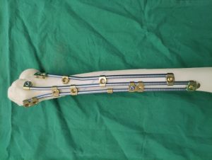
Pic : Ortho-Bridge System (OBS)
In severe trauma instances, the surgeon conducting the intramedullary nailing should be aware that respiratory difficulty might occur. They should also be aware of the various nail sizes available at their institution, as technique and equipment alter depending on the size of the nail. Reamers extend the medullary canal in most situations to allow for large-diameter nails. Cannulated or solid nails are available. Solid nails, due to their absence of internal dead space, may help to save the patient against infections that might occur during or after the treatment. The diameter of the nail or screw increases the endurance and strength of the nail or screw.
Reaming has the drawback of causing harm to the medullary blood supply. As a result, the diaphysis of the bone suffers from transient necrosis. Although this scenario may have a higher chance of infection, multiple clinical research has demonstrated that this is not a major risk. Sharp reamers should be cut through bone with the least amount of friction feasible. The flutes should be deep, and the shaft should be tiny in diameter. The major goal is to reduce the pressure in the canal and avoid friction that would cause the bone and soft tissues surrounding it to overheat.
Intramedullary nail fixing is a difficult yet simple operation. The medullary tube is passed through the fracture site and over the reaming guide wire. The reaming guide is then retrieved while it is kept in place. The nailing wire is inserted into the tube, and its location is verified using x-rays. After that, the medullary exchange tube is removed.
Reaming is still preferred by most surgeons; however, it should be done with extreme caution when the canal width is small, and the diaphysis of the radius or ulna is thick. Because the nails fit tightly in the more extended place of the reamed process, it provides better stability. Non-reamed nails were made to cut down on the risk of infection and respiratory problems caused by medullary debris that had been embolized [68]. It’s proven to be beneficial in medullary canals in small diameter. However, the therapeutic advantages are negligible since non-reamed nails with tiny diameters are weak. As a consequence, it has a greater reoperation rate.